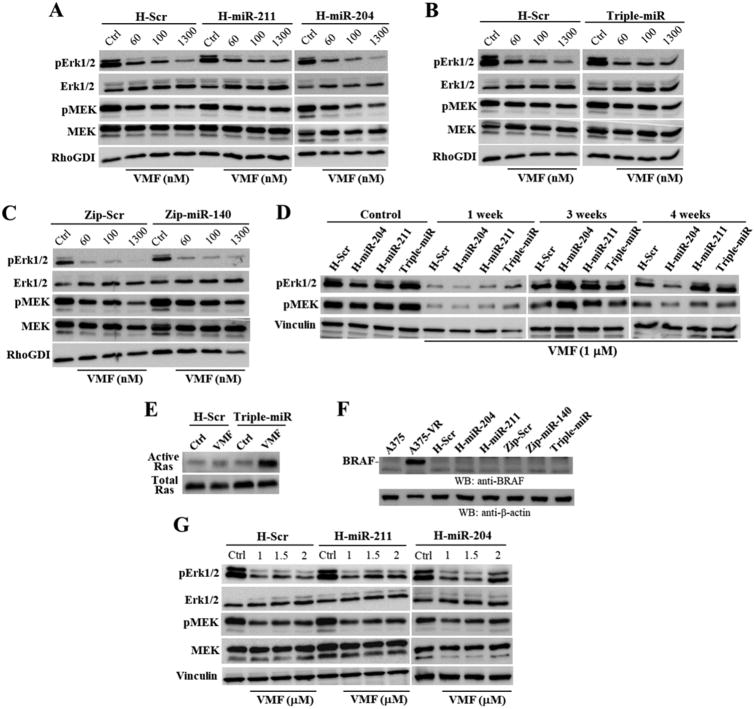
Figure 6. Analysis of Erk1/2 and MEK activation in miRNA transductants.
(A-C) A375 transductants were incubated for 24 h in the absence (Ctrl) or presence of the indicated concentrations of VMF, and subsequently subjected to immunoblotting. (D) Cells were left untreated (Control) or incubated for the times shown with VMF, and analyzed by western blotting. (E) Cells were incubated for 24 h without (Ctrl) or with VMF (1.3 μM) and subsequently subjected to Ras GTPase assays. (F) Parental and A375-VR cells, or the indicated transductants were analyzed by immunoblotting using anti-BRAF antibodies. (G) SK-Mel-28 transductants were incubated for 24 h with or without VMF, and subsequently subjected to immunoblotting. Loading controls were assessed with antibodies to RhoGDI, vinculin or β-actin.

