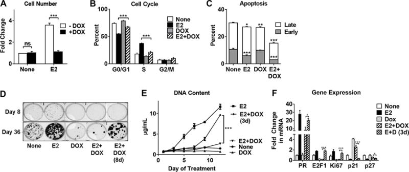
Figure 2. CA-IKKβ reversibly inhibits E2-induced proliferation and enhances cell survival.

A-C, MCF-7-CA-IKKβ cells were treated with E2 (10 nM), DOX (1 μg/mL), or E2+DOX for 72 hrs. Cells were trypsinized and counted using a hemocytometer (A), fixed and stained with propidium iodide for cell cycle analysis (B), or fixed and stained with Annexin V-FITC and propidium iodide to assess apoptosis (C). D, MCF-7-CA-IKKβ cells were seeded as single cells and treated with E2, DOX, or E2+DOX for 36 days. On day 8, DOX was withdrawn from one E2+DOX group. Colonies were visualized following methanol fixation and crystal violet staining. E, MCF-7-CA-IKKβ cells were treated with E2, DOX, or E2+DOX for 12 days. After 3 days, DOX was withdrawn from one E2+DOX group. Cells were retreated every 2-3 days. DNA content was determined by Hoechst 33342 staining. F. To assess reversal of gene expression, MCF-7-CA-IKKβ cells were treated with E2, DOX, or both for 8 days and DOX was withdrawn from one group on day 3. RNA was isolated and gene expression was assessed by RT-QPCR for E2-regulated genes. (* P<0.05, ** P<0.01, *** P<0.001 and ns=non-significant).
