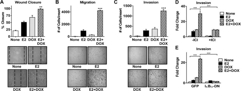
Figure 4. E2 and CA-IKKβ work together to promote migration and invasion.

A. MCF-7-CA-IKKβ cells were grown to ~80% confluence and a wound was created. Cells were then washed and treated with E2, DOX, or E2+DOX for 96 hrs. Wound closure was measured as a percent of initial wound size (** P<0.01 vs all other groups). B, C. MCF-7-CA-IKKβ cells were treated with E2, DOX, or E2+DOX for 72 hrs. Equal numbers of cells were transferred to migration (B) or invasion (C) transwell inserts and allowed to migrate or invade for 24 hrs. Cells were fixed, stained and counted manually. (*** P<0.001 vs all other groups). D. MCF-7-CA-IKKβ cells were treated with E2+DOX in the presence or absence of 1 μM ICI 182,780 for 72 hr prior to conducting an invasion transwell assay. (***P<0.001). E. MCF-7-CA-IKKβ cells were infected with adenovirus expressing a dominant negative form of IκB (IκBα-DN) or GFP as a control prior to treatment and an invasion transwell assay. (***P<0.001).
