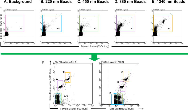
Figure 2. Detection of polysterene microparticles to determine flow cytometer detection limit.
(A) PBS buffer alone that has been filtered and ultracentrifuged used to define instrument background noise. The variously colored R1 gate excludes background noise (B) 220 nm polysterene beads diluted in cleared PBS buffer can be distinguished from background noise based on Side Scatter profile. (C) 450 nm polysterene beads diluted in cleared PBS buffer can be distinguished from background noise based on Side Scatter profile. (D) 880 nm polysterene beads diluted in cleared PBS buffer can be distinguished from background noise based on Side Scatter and Forward Scatter profiles. (E) 1340 nm polysterene beads diluted in cleared PBS buffer can be distinguished from background noise based on Side Scatter and Forward Scatter profiles. (F) Combination of 220, 450, 880, and 1340 nm beads detected in the Green Fluorescence channel v. Forward Scatter channel and Green Fluorescence channel v. Side Scatter channel. Different sized particles are outlined with gates corresponding to their R1 gate. Millipore’s Guava easyCyte 8HT second generation flow cytometer was used with Spherotech’s SPHERO™ Nano Fluorescent Particle Size Standard Kit Beads (Cat No. NFPPS-52-4K).

