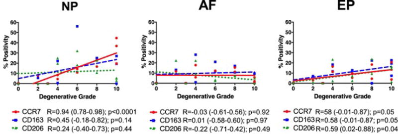
Figure 5. Percent positivity of macrophage phenotype markers significantly increased in NP and EP but not AF regions.

The percentage of cells immunopositive for CCR7, CD163, and CD206 were averaged within NP, AF, and EP regions for individual IVDs and correlated with Rutges degenerative grade. CCR7+, CD163+ and CD206+ cells all significantly increased with degenerative grade in endplate (EP) regions while only CCR7+ cells significantly increased in NP regions. No changes in percent positivity of any macrophage marker were observed with degenerative grade in AF regions. (N = 12). R = Spearman’s correlation coefficient (95% Confidence Interval).
