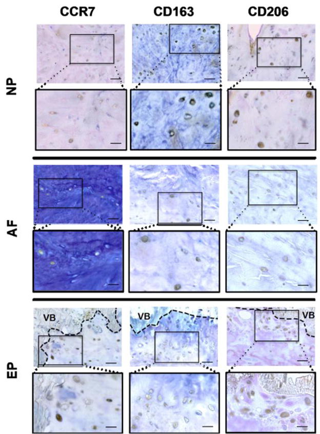
Figure 7. Macrophage phenotype markers were observed in unhealthy areas in the NP, AF, and EP regions of IVDs with moderate and high degeneration.

Unhealthy areas were identified as irregularities and obvious defects in extracellular matrix organization and cellular distribution patterns including proximity to tears, cracks and ruptures, granulation-like tissue, irregularities in collagen fiber orientation, cell clustering and cells with irregular size, shape or organization pattern. Scale bar (in unboxed images) = 50 μm, (in boxed images) = 25 μm. VB = vertebral bone.
