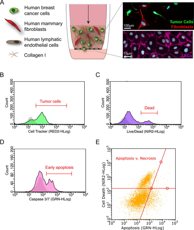
Figure 5. Design and analysis of a human 3D in vitro model of the breast tumor microenvironment.

A) Schematic of a physiologically relevant human 3D in vitro model of the breast tumor microenvironment. Fluorescently-labeled human breast tumor cells and human mammary fibroblasts are incorporated in a Collagen I matrix seeded atop the porous membrane (8 μm pore size) of a tissue culture insert (inset-top: fibroblasts (red), tumor cells (green), nuclei (DAPI-blue). A confluent monolayer of human lymphatic endothelial cells is seeded on the alternate side of the membrane (inset bottom: CD31 (pink), nuclei (DAPI-blue)). B) Flow cytometry is used to identify (B) Cell Tracker-labeled breast tumor cells (C) uptake of live/dead indicator to assess cell death (D) Caspase 3/7 positivity to indicate apoptosis and (E) Track total cell death via apoptosis and necrosis. Negative controls are shown in gray on histogram plots.
