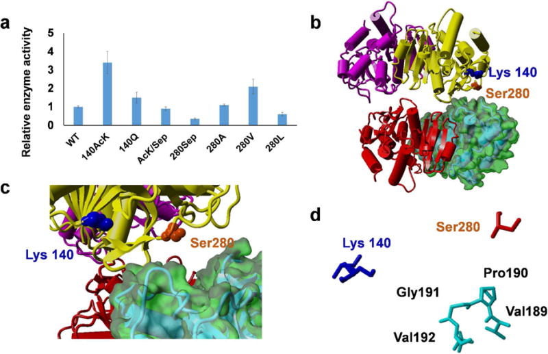
Figure 5. The structure and function of MDH.

A) The relative enzyme activities of MDH and its variants. Mean values and standard deviations were calculated from three replicates. The enzyme activity of wild-type MDH was set as 1. B) The crystal structure of E. coli MDH tetramer (PDB ID: 3HHP). C) A zoom-in view of the dimer-dimer interface. D) A map of amino acids involved in the dimer-dimer interface.
