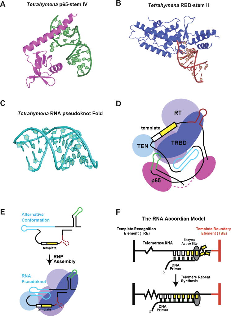
Figure 2. RNA structure and function in ciliate telomerase.
(A) High-resolution structure of the Tetrahymena p65 protein xRRM protein domain bound to stem-loop IV of telomerase RNA. Figure adapted from (36) PDB 4ERD. (B) High-resolution structure of the Tetrahymena TERT-RBD domain bound to the base of stem-loop II, comprising the template boundary definition complex. Figure adapted from (37) PDB 5C9H. (C) High-resolution structure of the Tetrahymena RNA pseudoknot domain. Figure adapted from (38) PDB 5KMZ. (D) Schematic model of Tetrahymena telomerase RNA organization based upon the medium-resolution structure of the complete holoenzyme solved by cryo-electron microscopy. Figure adapted from (39). (E) Cartoon model for reorganization of Tetrahymena RNA pseudoknot fold upon binding and assembly with TERT protein subunit. (F) The RNA accordion model for RNA structural rearrangements during telomerase catalysis. During telomere DNA repeat synthesis the RNA regions flanking each side of the template undergo compaction and expansion to facilitate movement of the template through the RT active site.

