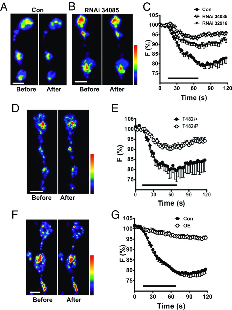
Fig. 3.
Myopic regulates activity-induced synaptic neuropeptide release. Pseudocolor images of synaptic boutons expressing Dilp2-GFP in control animals (Con) (A) and Myopic RNAi (no. 34085) knockdown (B) before and after 70-Hz stimulation for 1 min. (C) Time-course DCV release in response to 70-Hz stimulation (indicated by bar) in Con and two lines of Myopic RNAi knockdown (RNAi, nos. 34085 and 32916) in synaptic boutons expressing Dilp2-GFP [Con (five animals); RNAi no. 34085 (12 animals), and RNAi no. 32916 (11 animals)]. F, fluorescence. (D) Pseudocolor images of heteroallelic (T482/P) Myopic mutant NMJs expressing Dilp2-GFP before and after 70-Hz stimulation. (E) Time-course DCV release in T482/+ (four animals) and Myopic mutant boutons (T482/P, nine animals) expressing Dilp2-GFP. (F) Pseudocolor images of boutons labeled with Dilp2-GFP overexpressing (OE) Myopic before and after stimulation. (G) Overexpression of Myopic (five animals) inhibits neuropeptide release in synaptic boutons (Con, six animals). (Scale bars, 2 μm.)

