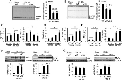
Fig. 2.
Changes in expression of cleaved caspase-8, RIP1, RIP3, and MLKL in the kidneys of WT and Rgmb cKO mice after IRI. Male WT and Rgmb cKO mice at 2 mo of age underwent 40 min of bilateral renal pedicle clipping. Mice were killed after 12 and 24 h. (A and B) Cleaved caspase-8 levels in kidneys 12 and 24 h after IR. Kidney lysates 12 (A) and 24 h (B) after reperfusion were subjected to Western blotting using an antibody that recognizes both full-length and cleaved caspase-8 (Casp-8). Cleaved caspase-8 levels relative to full-length caspase-8 levels were quantified by densitometry. (C–E) mRNA levels of Rip1, Rip3, and Mlkl in kidneys 12 and 24 h after IR. Kidneys collected from WT and cKO mice subjected to sham operation or IR were analyzed for mRNA levels of Rip1 (C), Rip3 (D), and Mlkl (E) by real-time PCR. (F and G) Protein levels of RIP3 and MLKL in kidneys 12 and 24 h after IR. Kidneys collected from WT and cKO mice subjected to sham operation and IR were analyzed for protein levels of RIP3 (F) and MLKL (G) by Western blotting. RIP3 and MLKL levels relative to GAPDH levels were quantified by densitometry. Rpl19 was used as the internal control for real-time PCR. GAPDH was used as the loading control for Western blotting. n = 5–6 for C–E. *P < 0.05; **P < 0.01; ***P < 0.001.

