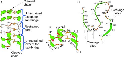
Fig. 3.
Fibril template used in the MD simulations. The initial template conformation is taken from the model of Xiao et al. (24). (A) An Aβ1–42 fibril template, consisting of six chains, is used to represent a fibril. Backbone restraints are used to stabilize the two chains at the core of the fibril (depicted in white). The two chains around the core are not restrained, except to stabilize the salt bridge between K28 and A42, which is critical for the stability of the structure (Fig. 2C). Only the chains at the two ends of the template (the top and bottom layers in the diagram) are cleaved. (B) The layers of a fibril are held together by intermolecular hydrogen bonds; their position and direction are indicated by red arrows. Hydrogen bonds along the β-strand regions are expected to be more stable compared with the turn-and-coil regions. Only hydrogen bonds along the β-strand regions are indicated in the diagram. (C) A single layer of an Aβ1–42 fibril showing the eight cleavage sites used in our simulations. For each simulated system only one cleavage site was used, with the cleavage applied at both the top and the bottom layer simultaneously.

