
Fig. 2.
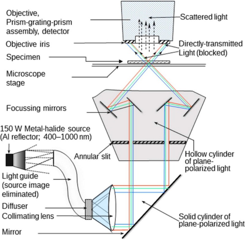
The various components in a CytoViva HSI system [14]. Briefly, the components are (i) a light source; (ii) a high resolution light collimator or adapter; (ii) mirror(s) to generate plane-polarized light or light dispersive elements to disperse incident white light into its constitutive spectra; (iii) a microscopy stage for the specimen; (iv) an optical module for bright- and dark-field analysis; (v) visible and near infra-red spectrometer to collect and convert electromagnetic energy into electrical signals for image formation; (vi) an image capture modality (see Fig. 3) and (vii) computer for data collection and software analysis (not shown).