
Figure 1.
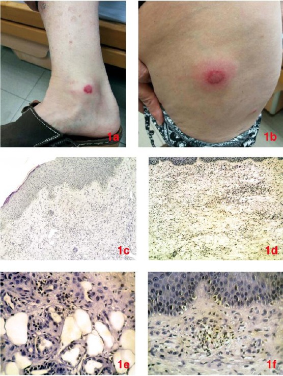
Nodular skin lesions localised at legs (a) and thighs (b). Incisional biopsy revealed normal epidermis and dense inflammatory infiltrate in the dermis (c), mainly composed of lymphocytes and histiocytes (d) [Haematoxylin and eosin stain, x40]. The infiltration extended to the subcutaneous adipose tissue (e), showing a pale neutrophilic background (f) [Haematoxylin and eosin stain, x100]