
Figure 1.
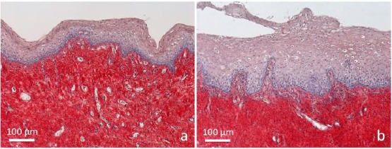
Samples of vaginal mucosa respectively a) before and b) 1 hour after laser application. a) The epithelium does not present any superficial desquamation, and its basal surface appears relatively smooth. The connective tissue is intensely stained; b) the epithelium is thicker, formed by bigger cells distributed in very numerous layers. At the surface, it appears desquamating in the vaginal lumen. The connective tissue is penetrating into epithelial indentations as deep bell-shaped structures constituting newly formed papillae. Papillae are formed by a loose connective tissue with small and thin fibrils inside, faintly stained by Picrosirius red. Many penetrating small vessels, are observable inside them. Light microscopy (not polarised light), sections 5 µm thick. Picrosirius red and fainty Hematoxylin staining