
Figure 2.
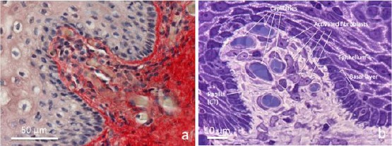
Connective tissue bell-shaped papillae in vaginal mucosa samples 1 hour after laser application. a) Paraffin section (5 µm) stained with Picrosirius red and light Hematoxylin, b) semithin section (0.2 μm) from resin embedded specimen, stained with toluidine blue. a) at the centre towards the high left, the connective tissue of papilla seem “pushing” underneath the epithelium by the growing connective tissue rich in vessels. In the loose and faintly stained connective tissue of papilla, very thin red fibrils between blood capillaries are observable. b) The semithin section permits high-resolution light microscopy. Transverse sections of blood capillaries, small pericytes around endothelium and fibroblasts with clear nuclei and well visible nucleoli, and basophilic cytoplasm are observable. Into the epithelium facing the connective tissue, the basal layer appears organised by a single row of cuboidal cells with basophilic cytoplasm. Light microscopy (not polarised light)