
Figure 3.
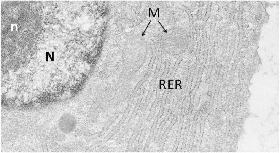
Electron micrograph representing a papillary fibroblast. Part of the nucleus (N) rich in euchromatin is showing inside a compacted nucleolus (n). In the cytoplasm, a highly extended rough endoplasmic reticulum (RER) rich in ribosomes, containing inside cisternae a finely filamentous material, is well visible. M: mitochondria