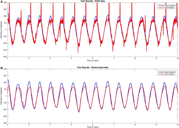
Figure 1.
RAW and desaccaded visually enhanced VOR (VVOR) plots. (A) Original (RAW) VVOR plot from a patient with a unilateral vestibular hypofunction as a consequence of a retrolabyrinthine left vestibular nerve section performed due to a vestibular schwannoma. (B) Desaccaded eye curve and plot. Eye velocity plots were inverted to visually match the head velocity plots.

