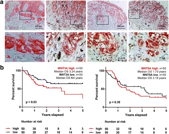
Fig. 1.

β-catenin is expressed in primary human melanomas in particular in cells of the invasive front. a Cytoplasmic expression of β-catenin in primary melanomas. A stronger expression of β-catenin in melanoma cells of the invasive front was observed by immunohistochemistry when compared to the bulk cells of the primary melanoma. Melanoma cells of the invasive front with a high expression of β-catenin had a spindle-like, mesenchymal morphology. Overview (upper row) and higher magnification of the invasive front (lower row) of four representative primary melanomas. b Overall survival (OS) analysis of the TCGA data set for the top and bottom (n = 50 each) WNT3A and WNT5A expressing melanomas reveal a significantly worse OS for WNT3A-high (p = 0.03) but not WNT5A-high melanoma patients (p = 0.39)
