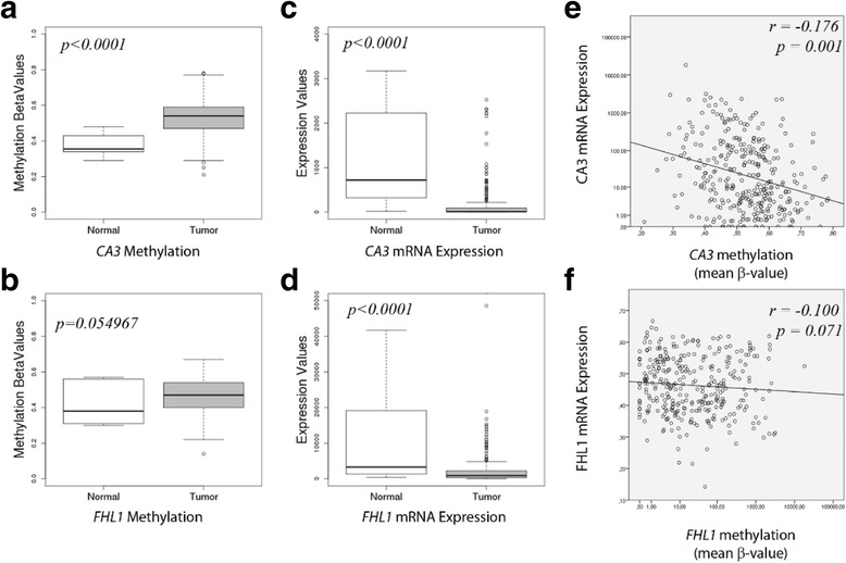
Fig. 4.

In silico TCGA evaluation of methylation status and mRNA expression of CA3 and FHL1. a and b boxplots show mean methylation β-values in normal and tumor tissues, for CA3 and FHL1 genes, respectively. c and d boxplots show mRNA expression distributions, in normal and tumor tissues, for CA3 and FHL1 genes, respectively. Statistical p values denote Student’s ttest between normal and OSCC samples. e and f show Pearson’s correlation of mean methylation β-values and mRNA expression, for CA3 and FHL1 genes, respectively
