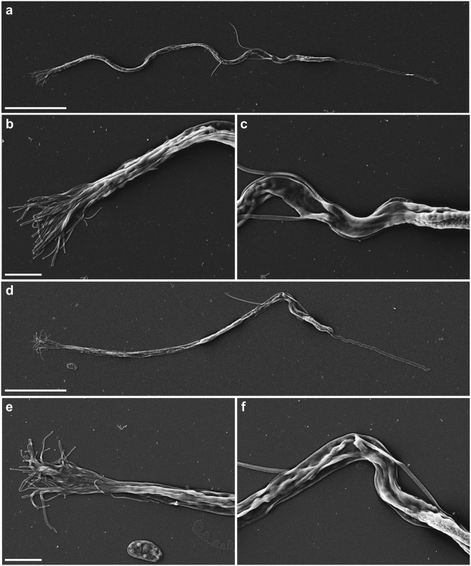
Figure 2.
Scanning electron microscopy of spermatozoa. (a–c) Spermatozoon of a negative control GFP(RNAi) worm. (a) Overview of the complete cell. Note the curved view of the shaft. (b) Detail of the brush, consisting out of separate extensions. (c) Detail of the notch region of the cell. (d–f) Spermatozoon of a Mlig-sperm1(RNAi) worm. (d) Overview of the complete cell. Note the rigidity of the shaft and the contortion at the notch site. (e) Detail of the brush. Compared to the negative control the brush looks more flattened with the base of the extensions being more packed together. (f) Detail of the notch region clearly showing the contortion. Scale bar are 10 µm (a,d) and 2 µm (b,c,e,f).

