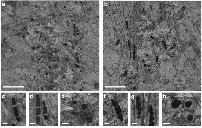
Figure 3.
Ultrastructure of spermatids and spermatozoa. (a,b) Overview of early and late spermatids and spermatozoa in the testes of a negative control GFP(RNAi) worm (a) and a Mlig-sperm1(RNAi) worm (b). In both images, several longitudinal and cross sections of nuclei of the spermatozoa can be observed as black structures. (c,d) Detail of longitudinal sections of spermatozoa nuclei of a GFP(RNAi) worm. The chromatin of the nucleus is condensed into discrete bodies. (e) Detail of a cross section of spermatozoa nuclei of a GFP(RNAi) worm. (f,g) Detail of longitudinal sections of spermatozoa nuclei of a Mlig-sperm1(RNAi) worm. Compared to the negative control, the chromatin of the nuclei looks fragmented and condensation into discrete bodies is less visible. (h) Detail of a cross section of spermatozoa nuclei of a Mlig-sperm1(RNAi) worm. Compared to the negative control, the chromatin of the nuclei is more fragmented. Scale bars are 5 µm (a,b) and 500 nm (c–h).

