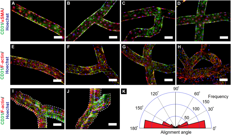
Figure 2.
Formation and characterization of microvascular network. (A–H) Lumenized vasculature developed by human breast tumor-associated endothelial cells (hBTECs) maintained under flow in various sections of the microfluidic channels; cells displayed an elongated morphology with a high degree of vascular maturity and cell-cell contact. (A–D) (CD31, green; αSMA, red; nuclei, blue) and (E–H) hBTECs (CD31, green; F-actin, red; nuclei, blue) (I–J) 3D projected views of vascular channels exhibited complete cellular coverage of the channels (white lines indicate edges of microchannel). Scale bar = 100 μm. Vascular flow is oriented from left to right and from top to bottom in the images. (K) Circular histogram of hBTEC orientation when maintained under continuous flow. A high degree of cellular alignment with respect to flow direction was observed in the channels (n = 444 cells from over 10 separate locations on each of three independent chips).

