Figure 5.
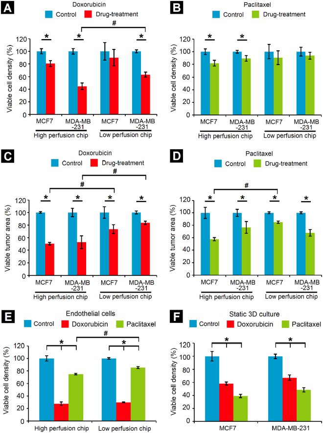
Drug-testing in tumor-mimetic chips. Reduction in viable cell density due to (A) Doxorubicin and (B) Paclitaxel. Viable MDA-MB-231 cell density (co-encapsulated with fibroblasts) following doxorubicin treatment was significantly lower in the HPC design as compared to the LPC design; this trend was not observed for the MCF7 cells (co-encapsulated with fibroblasts). No significant differences in viable cell density in either chip design were observed following paclitaxel treatment. Reduction in viable tumor area (area occupied by viable cells) due to (C) Doxorubicin and (D) Paclitaxel treatment revealed that doxorubicin caused a significant reduction in viable tumor area for both the cell lines in the HPC design as compared to the LPC design. Paclitaxel treatment caused a significant reduction in viable tumor area of MCF7 cells but not MDA-MB-231 cells. (E) Drug cytotoxicity on endothelial cells demonstrates greater cytotoxicity of doxorubicin as compared to paclitaxel in both chip designs. (F) Decrease in viable cell density of both cell lines in static 3D well plate culture (*Significant difference between control and drug treatment groups, p < 0.05; #Significant difference between same cell type in different chip designs, p < 0.05, n = 3 independent chips per condition; for static 3D culture, n = 3 independent hydrogel constructs per condition, error bars represent standard deviation).

