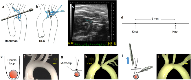
Figure 1.
(a) Rockman technique. (b to j) Schematic description of steps for the Double loop-clip technique. (c) B-mode echocardiographic image in modified right parasternal view of the thoracic aorta used to measure mid aortic arch luminal diameter, between the innominate trunk and the left carotid artery. This distance was obtained preoperatively and used to calculate the inter-knot span of the suture for the double loop-clip technique and customize the constriction to the mouse´s somatometry. (d) The suture was prepared prior to the operation with two knots spread apart a controlled distance (5 mm in the example) intended to define the degree of constriction. (e) The suture is looped twice around the mid-aortic arch (f), and (g) a vascular microclip applied under both knots which produces the preassigned level of stenosis as seen in the silicone cast of the thoracic aorta (h). (i) During the reoperation for releasing the constriction, pulling from the microclip allows its separation from the aortic wall, which facilitates severance of the suture avoiding the risk of aortic laceration. After release of the constriction the aorta recovers its flow section even though a mild stenosis remains (h). DLC: Double loop-clip.

