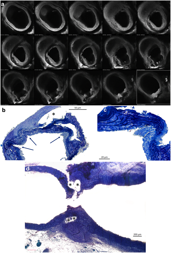
Figure 6.
Histological changes in the transverse aortic arch four weeks after TAC with the DLC technique. (a) Confocal microscopic images of the murine aortic arch. The transverse sections progress from proximal (upper left) to distal arch (lower right). As the section with the minimal luminal area (middle row, center panel) is approaching, a progressive thickening of the intimal layer is observed. This phenomenon does not appear distally to the constriction. Toluidin blue stained semithin sections of the aortic mid arch in the area of constriction (b to d). (b and c) Transverse sections disclose outward folds of the aortic wall in the vicinity of the constriction that (c) are partial or totally occupied by cellular elements, whereas the lamellar medial units appear compressed and devoid of smooth muscle cells. Adjacent to the folds the media appears reticulated and thickened with normal cellularity. (d) A residual empty channel with a fibrous sheath cover (*) reveals the position of the constricting suture in a longitudinal section.

