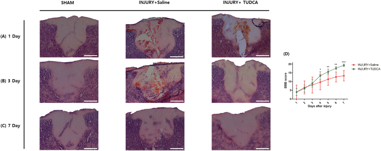
Figure 5.
Histopathological effect of TUDCA by H&E staining and behavior tests in injury and TUDCA group. Lesion epicenter histopathology (H&E staining): (A) Spinal cord tissue on 1 day after spinal cord injury. (B) Spinal cord tissue on 3 day after the injury shows more remaining neurons and low inflammation in the TUDCA group. (C) Spinal cord tissues on 7 day after the injury are similar in the TUDCA and sham groups (Scale bar = 200 µm). (D) The Basso-Beattie-Bresnahan (BBB) scores for the hindlimb function were evaluated for 7 days after the injury. The TUDCA group received intraperitoneal injections of 200 mg/kg of TUDCA at one minute and 24 hours after establishing the model. The injury group received intraperitoneal injections of the same dose of saline at the same time points. Results are the mean ± SD of triplicate experiments: *p < 0.05, **p < 0.01, and ***p < 0.001, significant difference as compared to the 1 day for each group by unpaired Student’s t-test.

