Figure 1.
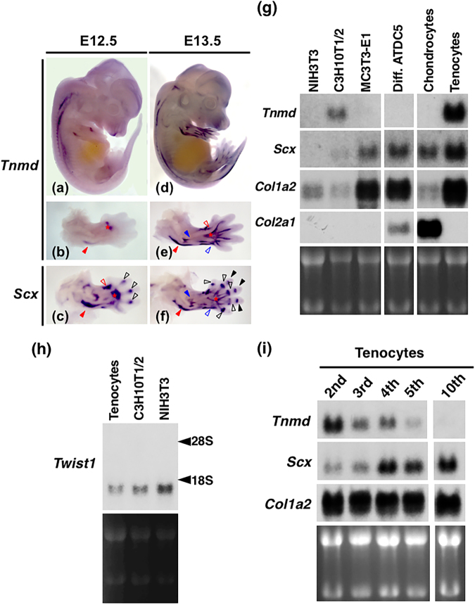
Coexpression of Tnmd and Scx in the developing tendons. (a–f) Expression patterns of Tnmd (a,b,d,e) and Scx (c,f) in embryos at E12.5 (a–c) and E13.5 (d–f) are shown. Whole-mount in situ hybridisation with antisense probes for these two genes were performed. Lateral (a,d) and dorsal views of the forelimbs (b,c,e,f) are shown, respectively. Red, black, and blue arrowheads indicate the developing triceps branchii tendon, joint capsules between proximal and middle phalanx, and extensor digitorum communis, respectively. Open red, black, and blue arrowheads indicate the developing extensor carpi radialis brevis/longus tendon, joint capsules between the metacarpus and proximal phalanx, and extensor carpi radialis tendon, respectively. Asterisks (b,c,e,f) indicate the extensor digitorum communis tendon. (g) Total RNA was prepared from NIH3T3 cells on day 3, C3H10T1/2 cells on day 3, MC3T3-E1 cells on day 14, differentiated ATDC5 cells on day 21 (Diff. ATDC5), primary rat costal chondrocytes on day 24 (Chondrocytes), and a secondary culture of rat limb tendon-derived tenocytes (Tenocytes). Detection of Tnmd, Scx, Col1a2, or and Col2a1 by northern blotting is shown. (h) Detection of Twist1 by northern blotting is shown. Arrowheads indicate the positions of ribosomal RNA subunits. (i) Total RNA was extracted from confluent 2nd, 3rd, 4th, 5th, or 10th passages of tenocytes isolated from rat leg tendons. Northern blot analysis of Tnmd, Scx, and Col1a2 is shown. Total RNA (15 µg) was loaded in each lane and the loading levels were verified by ethidium bromide staining.

