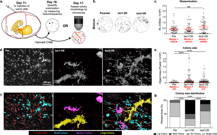
Figure 2.
Full length ALCAM (ALCAM-Iso1) mediates tumor cell cohesion. (a) Schematic describing avian embryo experimental metastasis assay. (b) Representative images of CAM colonization, measured by bioluminescence, following intravenous (IV) injection of indicated HT1080 cells. (c) CAM colonization was quantified using luciferase activity in a 1.5 cm core of CAM. Two cores per embryo from at least 10 embryos were measured. Relative bioluminescence (RL) represents mean value of two CAM sections per chick and was calculated using the integrated density tool (IntDen, 1 × 10⁶) in ImageJ. P-values were calculated using Kruskal-Wallis test with Dunn’s post-test, ns: not significant. (d) Representative images of metastatic colonies imaged 6 days post intravenous injection into CAM of chicken embryo. (e) Colony size was quantified using custom KNIME workflow. P-values were calculated using Kruskal-Wallis test with Dunn’s post-test, ****P < 0.0001. Graphs display mean with 95% confidence interval. Medians are reported. (f) Colonies were binned into single cell, small, medium, and large colonies using KNIME. Colonies are pseudo colored by size bin: Single Cells (Red), Small colonies (Cyan), Medium Colonies (Magenta), and Large colonies (Yellow). (g) Distribution of colonies across size bins was represented as percent of total area of all colonies. P-values were calculated using Chi-square test for trend, ****P < 0.0001. Graphs display mean with 95% confidence interval. Medians are reported.

