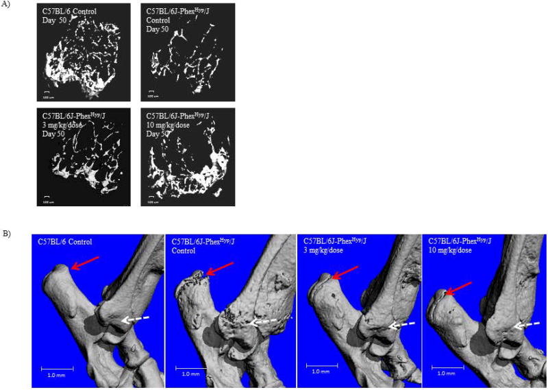
Figure 5. Improvement in bone mineralization and structure upon treatment of Hyp animals with 10mg/kg FGF23 c-tail Fc.
(A) Visualization of the cancellous bone of the distal femoral metaphysis as imaged by ex vivo µCT. A 100µm scale bar is indicated. (B) Depiction of bone quality as imaged via three-dimensional space filling analysis using µCT. For both these measures, these µCT images are representative of those in the following groups: C57BL/6 Control (n=10 mice); C57BL/6J-PhexHyp/J Control (n=10 mice); C57BL/6J-PhexHyp/J 3 mg/kg/dose (n=10 mice); C57BL/6J-PhexHyp/J 10 mg/kg/dose (n=10 mice). A 1mm scale bar is indicated.

