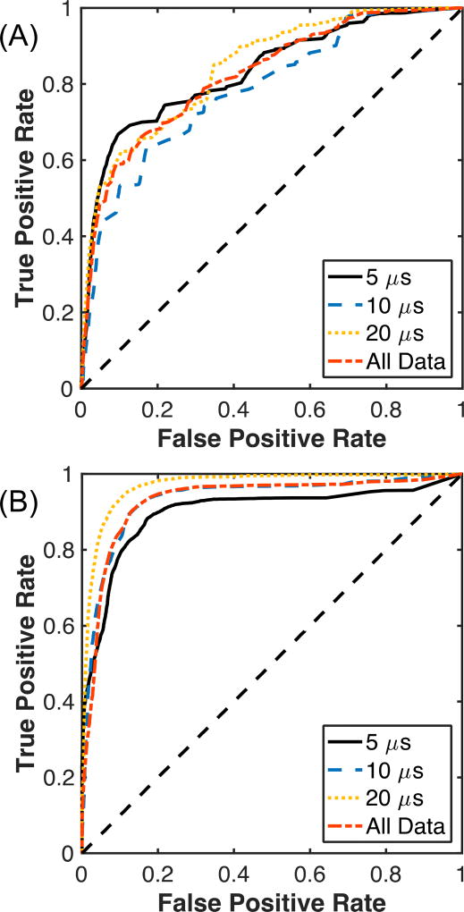
Fig. 4.
Receiver operating characteristic for (A) plane wave B-mode images and (B) passive cavitation images grouped by the histotripsy pulse duration, and for all pulse durations (All Data). No compression of the passive cavitation image acoustic power or grayscale values of the plane wave B-mode images was used in the computations of these values. N = 25 for each pulse duration.

