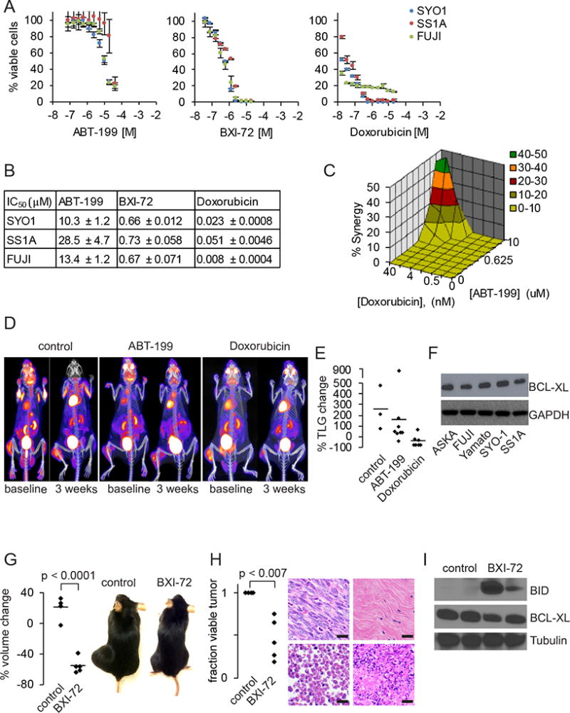
Figure 3. Targeting BCL2 and BCLxL in synovial sarcoma.

A. Cell viability curves for ABT-199, BXI-72 and doxorubicin in SYO-1 (blue), SS1A (red) and FUJI (green) after 48 hours of treatment.
B. IC50 data for ABT-199, BXI-72 and doxorubicin in SYO-1, SS1A and doxorubicin (± SEM).
C. Mac Synergy plot between doxorubicin and ABT-199 demonstrating minimal synergy (< 50% synergy) after 72 hours of treatment.
D. FDG-PET/CT image of Ptenfl/fl;hSS2 mice treated with control, ABT-199 or doxorubicin.
E. Graph of percent change in total lesion glycolysis (TLG) for synovial sarcomas treated with control, ABT-199 or doxorubicin.
F. Immunoblotting for BCLxL across five human synovial sarcoma cell lines with GAPDH as a loading control.
G. Graph depicting percent volume changes after 7 days of 20 mg/kg BXI-72 in Ptenfl/fl;hSS2 mice induced with local injection of TATCre and example gross photographs of Ptenfl/fl;hSS2 mice with synovial sarcomas on the left hindlimb treated with control DMSO or 20 mg/kg BXI-72.
H. Graph depicting percent necrosis in synovial sarcomas treated with control DMSO or BXI-72. Example H&E photomicrographs of necrosis identified in tumors treated with control DMSO or BXI-72: top left is viable tumor from control; top right is replacement of tumor with fibrosis in BXI-72 treated mice; bottom left is necrosis by ghost cells (anucleate); bottom right is necrosis/apoptosis by fragmentation.
I. Immunoblotting for BID (pro-apoptosis), BCLxL and tubulin in extracted synovial sarcomas treated with DMSO or BXI-72.
(All magnification bars = 10 µm)
