Fig. 1.
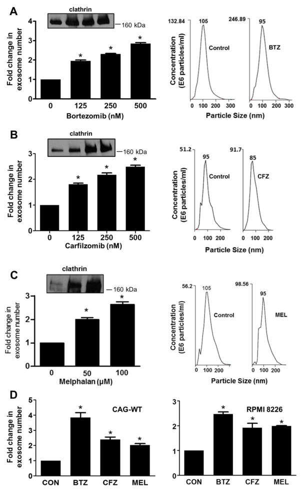
Chemotherapy enhances exosome secretion by myeloma cells. CAG human myeloma cells that express an elevated level of heparanase (CAG-HPSE) were treated with increasing concentrations of proteasome inhibitors bortezomib or carfilzomib (left panel, A, B) or the genotoxic drug melphalan (C). Exosomes released over 16 hours following exposure to drug were isolated by differential centrifugation and quantified by NanoSight particle tracking. The fold increase in the number of exosomes secreted following exposure of cells to drug is shown. Western blots for clathrin (Insets) of detergent extracts of exosomes secreted by an identical number of control and drug-treated cells (lanes 1–4 in A and B are 0, 125, 250 and 500 nM bortezomib or carfilzomib; lanes 1–3 in C are 0, 50 and 100 μM melphalan). In each case exosomes show increased amount of clathrin with increased drug concentration. Plots shown in the right panel, A, B are from NanoSight analyses of isolated exosomes from cells treated with 250 nM bortezomib or carfilzomib, in C cells were treated with 50 μM melphalan. The number shown at the peak of each plot is the median size of particles in that preparation. (D) CAG wild-type (CAG-WT) and RPMI 8226 human myeloma cells were treated with 250 nM bortezomib (BTZ), 250 nM carfilzomib (CFZ) or 50 μM melphalan (MEL) and the exosomes released over the 16 hours following exposure to drug were isolated by differential centrifugation and quantified by NanoSight particle tracking. The fold increase in the number of exosomes secreted following exposure of cells to drug is shown.

