Fig. 1.
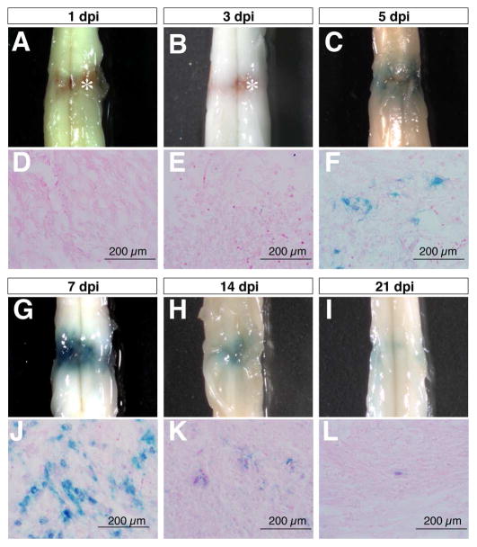
Compression spinal cord injury activates the Wnt signaling reporter TOPgal in a time-dependent manner. The wholemount spinal cords (A-C,G-I) and respective transverse sections (D-F,J-L) show the X-gal staining results for the TOPgal activities in the adult TOPgal mice at 1, 3, 5, 7, 14, and 21 days post injury (dpi). Note that the blue X-gal staining appears at 5 dpi (C,F), peaks at 7 dpi (G,J), and diminishes dramatically at 14 (H,K) and 21 dpi (I,L). Asterisks (A,B) indicate the reddish-brown substance caused by hemorrhage in the lesion site at 1 and 3 dpi. The sections were counterstained with Eosin solution.

