Figure 1.
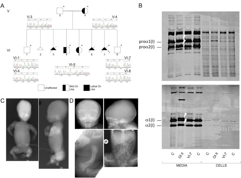
Homozygosity and heterozygosity for a 3bp deletion in CREB3L1 that results in a single amino acid deletion in OASIS (c.934_936delAAG [p.Lys312del]) are consistent with the variable severity of OI phenotypes. A) Pedigree of the CREB3L1 OI family. Arrow indicates the proband. Asterisks denote the family members whose exomes were sequenced. Chromatograms from Sanger sequence analysis confirming the exome findings are shown. B) Effect of the CREB3L1 variant on type I collagen biochemistry. Collagen alpha chains synthesized by skin fibroblasts from the proband (VI-7) are structurally equivalent to those from control fibroblasts (C), in contrast to those from a typical OI type II patient (OI II) with a COL1A1 glycine substitution (p.Gly818Cys), in which overmodification results in both delayed procollagen mobility (upper panel) (arrows) and delayed α1(I) and α2(I) chain mobility when procollagens are digested with pepsin (lower panel) (arrows). We do not see abnormal retention of procollagens nor of alpha chains in the proband fibroblasts; as skin fibroblasts do not express CREB3L1, we would not expect OASIS to have a role in regulating secretion in these particular cells. C) Anterior-posterior (left) and lateral (right) radiographs from VI-3 (homozygous for the 3bp deletion in CREB3L1) following 18 weeks gestation termination. There is virtually no calvarial mineralization. Bones in all visible extremities are telescoped and very irregular. The ribs are thin and wavy and there is mild platyspondyly. D) Radiographs of VI-4 (heterozygous for the 3bp deletion in CREB3L1) at birth. The calvarium is thin and the anterior and posterior fontanelles are large, but there are no Wormian bones. The ribs are thin and there is mild platyspondyly. There is marked bowing of the right femur.

