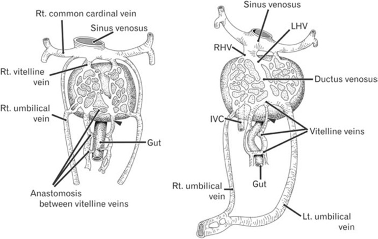
Fig. 1.
Diagram showing development of the portal and hepatic veins. The two vitelline veins communicate inside the liver and around the duodenum to form intrahepatic portal and hepatic veins: the left vitelline vein disappears, and the cranial part of the right vitelline vein and the segment that lies inferior to the liver give rise to the terminal branch of the IVC and portal and superior mesenteric veins. Incomplete involution and persistent communication of the vitelline venous system during the development of newly formed hepatic sinusoids results in various types of portosystemic shunts [11]

