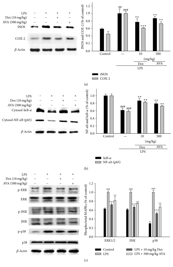
Figure 6.
The inhibition of iNOS, COX-2 (a), IκB-α, NF-κB (b), and MAPK protein (c) expression by AVA in LPS-induced ALI mice. Tissue homogenates were prepared and subjected to western blotting using antibodies specific for iNOS, COX-2, IκB-α, NF-κB, and MAPK. The values under each lane indicate the relative band intensities normalized to β-actin. The data are presented as the mean ± SD for three different experiments performed in triplicate. ###Compared with the control group. ∗∗ p < 0.01 and ∗∗∗ p < 0.001 compared with the LPS-alone group.

