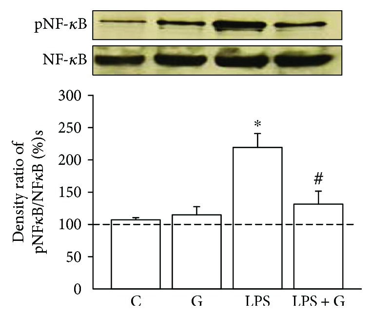
Figure 1.

G-CSF alleviated the elevated pNF-κB due to LPS treatment. Western blot analysis was used to detect the phosphorylated NF-κB (pNF-κB) p65 levels in the brain cell. The experimental groups are indicated as follows: vehicle-control (C), LPS group (LPS), G-CSF group (G), and LPS + G group (LPS + G) (n = 5 animals for each experimental group). The results were expressed as relative level of pNF-κB to total NF-κB. In the LPS group, the ratio of pNF-κB to total NF-κB was significantly higher compared to that in the LPS + G group and control group. ∗p < 0.05 compared with the control group. #p < 0.05 compared with the LPS group. Data are mean ± standard error of mean.
