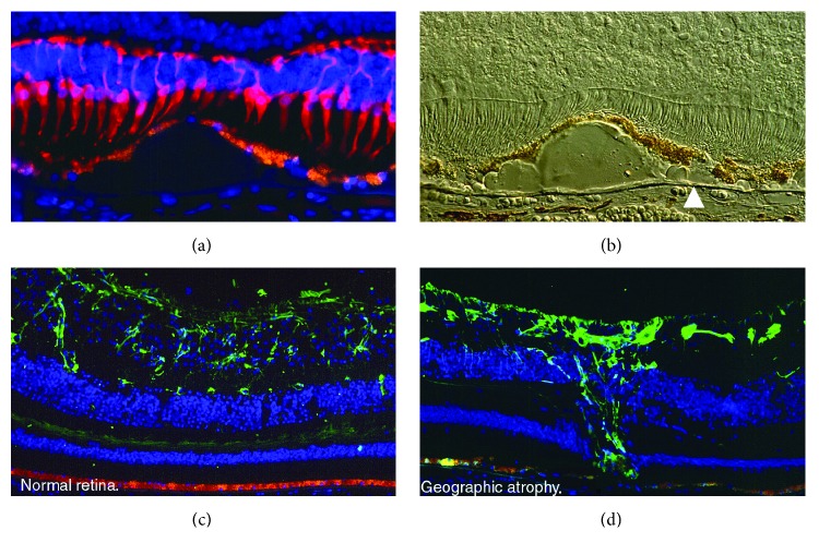
Figure 1.
(a) Representative immunofluorescence image of the macula with geographic atrophy and loss of cones (red cells, mAb 7G6) over drusen. The RPE (orange) is thinned over drusen. Cell nuclei are blue (DAPI). 40x objective. (b) Nomarski image of the previous image. Note refractile drusen on Brunch's membrane (arrowhead). 40x objective. (c) Representative immunofluorescence image of the macula in a normal retina. Orange (RPE) and green (GFP) in astrocytes (anti-GFAP). (d) Representative immunofluorescence image of the macula with geographic atrophy. Orange (RPE) and green (GFP) in Müller cell scar (anti-GFAP). Photo credit: “The Human Retina in Health and Disease” Teaching Set by Ann H. Milam Ph.D., University of Pennsylvania.

