
Fig. 1.
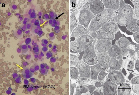
Bone marrow obtained before the initiation of treatment. a Bone marrow smear. Bone marrow is infiltrated by immature myeloma cells showing basophilic cytoplasm and fine nuclear chromatin networks with occasional nucleoli. Binuclear (yellow arrows) and trinuclear (black arrow) myeloma cells, suggesting hyperdiploidies, are seen (under × 40 magnification objective). b Electron micrograph of the bone marrow. Bone marrow is compactly occupied by immature myeloma cells with remarkable nucleoli, fine chromatin networks, and abundant rough endoplasmic reticula