Fig. 6.

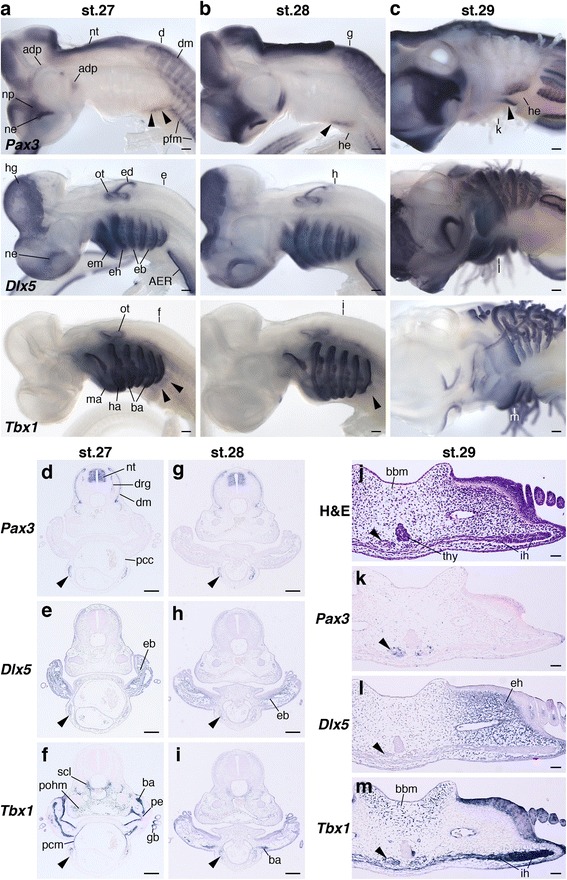
The arrangement of HBMs pathways and pharyngeal arches in shark embryos. Pax3, Dlx5 and Tbx1 expressions in sharks at stage 27 (a), 28 (b) and 29 (c). (a, b) Lateral and (c) ventrolateral views. Section in situ hybridization of Pax3, Dlx5 and Tbx1 at stage 27 (d–f), 28 (g–i) and 29 (k–m). Transverse section levels are at the fourth branchial arch (d–f), the second branchial arch (g–i) and the hyoid arch (j–m), as indicated in (a), (b) and (c). Arrowheads indicate HBM precursors. Pharyngeal arches and the pericardium were in the same transverse plane. Note that HBM precursors passed through the lateral side of pericardium, and then extended medially at the hyoid arch. Adp, anterodorsal lateral line placode; AER, apical ectodermal ridge; ba, branchial arch mesoderm; bbm, basibranchial mesenchyme; dm, dermomyotome; drg, dorsal root ganglion; eb, ectomesenchyme of branchial arches; ed., endolymphatic duct; eh, ectomesenchyme of hyoid arch; em, ectomesenchyme of mandibular arch; gb, gill bud; ha, hyoid arch mesoderm; he, heart; hg, hatching gland; ih interhyoideus; ma, mandibular arch mesoderm; ne, nasal epithelium; np, nasal prominence; nt, neural tube; ot, otic vesicle; pcc, pericardial cavity; pcm, pericardial mesoderm; pe, pharyngeal endoderm; pfm, pectoral fin muscle; pohm, postotic paraxial head mesoderm; scl, sclerotome; thy thyroid gland. Scale bars on whole embryos, 200 μm. Scale bars on sections, 50 μm
