Fig. 7.

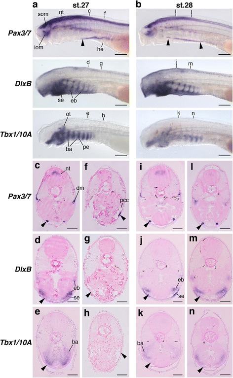
The arrangement of HBMs pathways and pharyngeal arches in lamprey embryos. Pax3/7, DlxB and Tbx1/10A expressions in lampreys at stage 27 (a) and 28 (b) from the lateral view. Transverse sections of stage 27 embryos at the fifth branchial arch (c–e) and heart (f–h) levels, and stage 28 embryos at the second (i-k) and fourth branchial arch (ln) levels. Section levels are indicated in (a) and (b). Arrowheads indicate HBM precursors. Pharyngeal arches and the pericardium were not in the same transverse plane, and HBM precursors expanded lateral to the pharyngeal arches. ba, branchial arch mesoderm; dm, dermomyotome; eb, ectomesenchyme of branchial arches; he, heart; iom, infraoptic muscle; nt, neural tube; ot, otic vesicle; pcc, pericardial cavity; pe, pharyngeal endoderm; se, surface ectoderm; som, supraoptic muscle. Scale bars on whole embryos, 200 μm. Scale bars on sections, 50 μm
