FIGURE 4.
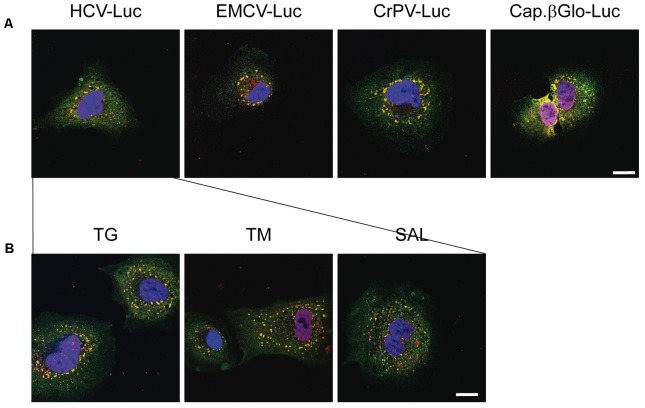
Thapsigargin, tunicamycin or salubrinal treatment induces stress granule formation in transfected Huh-7 cells. (A) Cells were seeded on microscope cover slips and transfected with different reporter RNAs: HCV-Luc, EMCV-Luc, CrPV-Luc or Cap.βGlobin-Luc. After 1 h of transfection, cells were incubated in DMEM medium for 2 h. (B) Cells were seeded on microscope cover slips and transfected with HCV-Luc. After 1 h of transfection, cells were treated with various eIF2 inhibitors (TG, TM, or SAL) for 2 h. In both cases, after treatments cells were permeabilized for immunocytochemistry using primary goat anti-TIA-1 and rabbit anti-eIF4G antibodies. An anti-goat antibody conjugated to Alexa 555 was used to detect TIA-1 (red) and an anti-rabbit antibody conjugated to Alexa 448 was employed to detect eIF4G (green). DAPI was used to stain the nuclei (blue). Scale bar, 20 μm.

