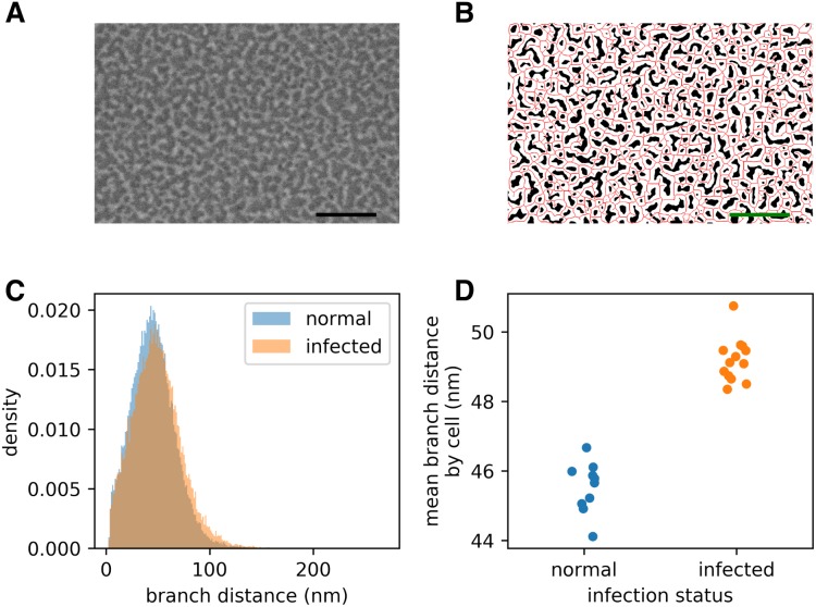
Figure 2. Infection by the malaria parasite remodels the spectrin skeleton of the host red blood cell in the asexual developmental stage.
(A) Example image produced by our protocol. Scale bar: 300 nm. (B) Thresholding (white) and skeletonisation (red) of the image in (A). (C) Complete distribution of measured spectrin branch lengths for normal and infected RBCs. (D) Mean spectrin branch length by cell (nnorm = 10, ninf = 13).

