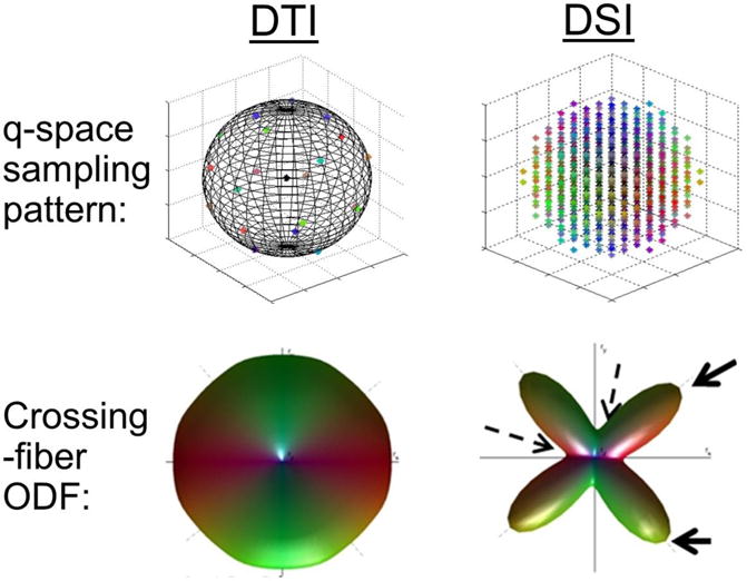
Figure 1.

Illustration of differences between diffusion tensor imaging and diffusion spectrum imaging (DSI), regarding its diffusion-space sampling (q-space) and orientation distribution function (ODF).
Note: In DTI, q-space is sampled on a spherical shell, whereas in DSI q-space is sampled on a Cartesian grid bounded by a spherical surface of given maximum b-value (typically much larger than usual b-values employed in DTI). In a simulation of a 90-degree, equal crossing fibers, DTI is unable to resolve the two directions, whereas the added samples in DSI allow for the computed ODF to resolve the two directions. The solid and dashed arrows point to the peaks and troughs of the ODF that are used to resolve diffusion directionalities.
