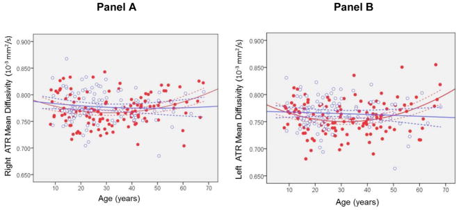
Figure 4. Mean diffusivity in the Right (Panel A) and Left (Panel B) Anterior Thalamic Radiation Modeled Using a Quadratic Curve Across the Agespan Separately in Males and Females.
Note: ATR=Anterior thalamic radiation. Dashed lines represent the 95% confidence intervals. Red dots are males and open blue circles are females. Units are 10−3 mm2/s.

