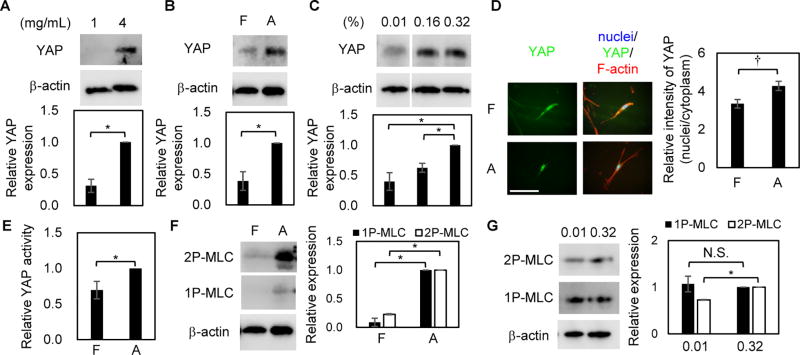
Figure 3. YAP and MLC are regulated by matrix stiffness in MSCs.
A, Western blot of YAP and β-actin in MSCs cultured with 4T1-conditioned media (CM) on floating collagen gels of 1 or 4 mg/mL collagen. n = 3 experiments. B, Western blot of YAP and β-actin in MSCs cultured with CM on floating (F) or attached (A) collagen gels of 1 mg/mL collagen. n = 4 experiments. C, Western blot of YAP and β-actin in MSCs cultured with CM on polyacrylamide gels of 0.01, 0.16, 0.32% BIS concentration. n = 3 experiments. D, Fluorescence images of nuclei, YAP, and F-actin in MSCs. Relative intensity of YAP in nuclei was quantified in n = 30 cells in 3 experiments. E, Luciferase assay of a YAP reporter construct in MSCs. n = 3 experiments. F, Western blot of 2P-MLC, 1P-MLC, and β-actin in MSCs. n = 3 experiments. G, Western blot of 2P-MLC, 1P-MLC, and β-actin in MSCs cultured with CM on polyacrylamide gels of 0.01 and 0.32% BIS concentration. n = 3 experiments. Statistical significance was determined as Supplementary Materials and Methods. N.S.: no significance with 95% confidence interval. Mean±S.E. are shown. Bar = 100 µm. MSCs were established from BALBc mice.

