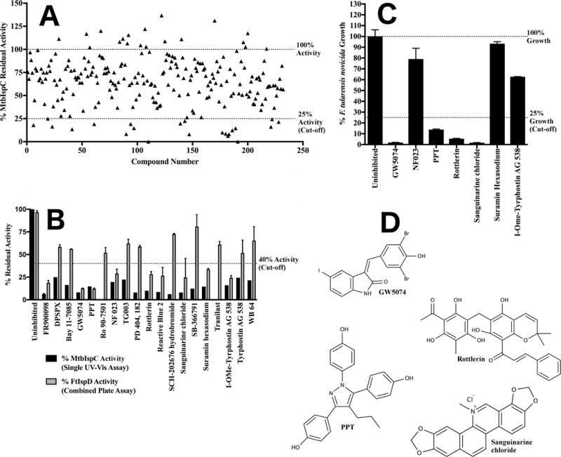
Figure 3. Screening of the LOPAC1280 library against IspC.

A) Secondary screening of the LOPAC1280 primary screen hits for IspC. Residual activity of MtbIspC when assayed in the presence of 100 μM of each of the LOPAC1280 library compounds identified in the primary screen. For clarity, each library molecule was assigned a numeric designation, ranging from 1–232 (the association of number, compound name, and residual enzyme activity is tabulated in Supplementary Figure 1B). Residual activity exceeding 100% reflects additional oxidation of NADPH over a well containing vehicle only. Library compounds demonstrating 75% or greater reduction in MtbIspC activity were selected for the tertiary screen. B) Tertiary screening of the LOPAC1280 library secondary screen hits for IspC via MtbIspC-FtIspD coupled assay. The residual activity of MtbIspC was measured with a coupled assay utilizing M. tuberculosis IspC and F. tularensis IspD (grey), and compared to the secondary screen values measuring MtbIspC activity using the A340 (black). Those compounds with less than 40% residual activity in the coupled assay were retained for quaternary antibacterial screening. Error bars are calculated as the deviation from the mean of two independent measurements. C) Quaternary screening of the LOPAC1280 tertiary screen hits for IspC against F. tularensis novicida. Antibacterial activity of LOPAC1280 inhibitors against F. tularensis subsp. novicida Utah 112 was measured in duplicate at 100 μM; compounds with less than 25% fractional bacterial growth were selected for EC50 determination. Fractional growth is calculated as the ratio of cell density (OD600) in the presence of 100 μM inhibitor to cell density in the presence of vehicle only. Error bars are calculated as the deviation from the mean of two independent measurements. D) Structures of lead compounds.
