
Figure 2.
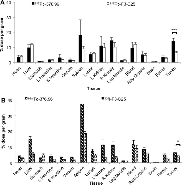
Biodistribution analyses in mice with PDAC xenograft tumors. (A) Biodistribution at 24 h after i.v. injection of 212Pb-376.96 or 212Pb-F3-C25 (0.74 MBq) in groups of athymic nude mice (n = 4/group) bearing s.c. PDX Panc039 tumors. (B) Biodistribution at 24 h after i.v. co-injection of 99mTc-376.96 (6.02 MBq) and 125I-F3-C25 (22.39 kBq) in athymic nude mice (n = 5 mice) bearing orthotopic PDAC3 tumors; 125I was counted after 99mTc had fully decayed. After euthanization of mice, tissues were collected, weighed, and counted to determine the percent of the injected dose per gram (% ID/g) of tissue. Data are presented as mean ± standard deviation. *p<0.05; ***p<0.001