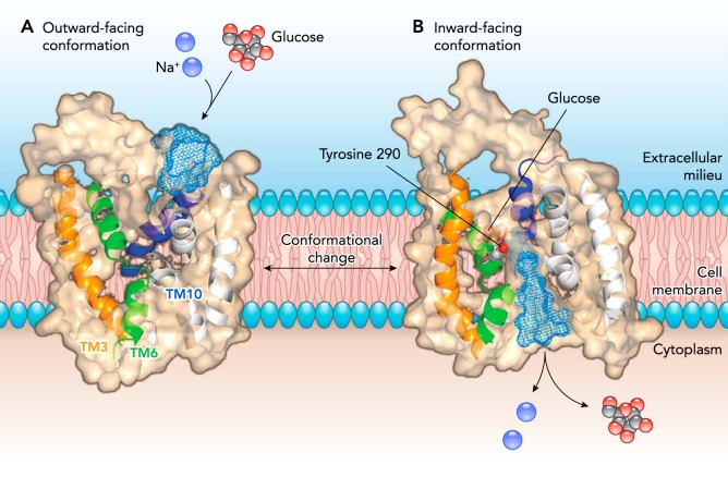
FIGURE 1.
Model of SGLT1 in the outward and inward facing conformations
In the open outward-facing conformation (left), a slice through the protein shows the opening to the external aqueous vestibule. External sodium binding to two Na+ binding sites (Na1 and Na2) causes the outer gate to open to allow external glucose to bind in the middle of the protein. A slice through the inward-facing protein (right) shows the bound glucose and the inward vestibule, through which Na+ and glucose escape into the cytoplasm. Helices showing structural rearrangements between the outward and inward conformations are shown in orange (TM3), green (TM6), and blue (TM10). Figure is adapted from Ref. 12, with permission, where the outward conformation is modeled on LeuT and the inward conformation is modeled on vSGLT.

